

[Sep-2025] Exam Sure Pass ARDMS Certification with SPI exam questions
Real ARDMS SPI Exam Questions Study Guide
NEW QUESTION # 71
What is a potential negative consequence of using a high wall filter?
- A. Aliasing could occur
- B. Desired signal may be eliminated
- C. Too much noise may appear on the image
- D. Penetration is reduced
Answer: B
Explanation:
A high wall filter is used in Doppler ultrasound to eliminate low-frequency signals that may be attributed to vessel wall motion or other low-velocity flows. However, if the wall filter is set too high, it can inadvertently eliminate desired low-frequency Doppler signals that represent real blood flow, particularly in smaller vessels or those with slower flow velocities. This results in a loss of valuable diagnostic information.
Reference: ARDMS Sonography Principles and Instrumentation (SPI) Review, Doppler Ultrasound section.
NEW QUESTION # 72
What is the result of increased transducer damping?
- A. Improved axial resolution
- B. Increased sensitivity
- C. Longer pulse length
- D. Narrow beam width
Answer: A
Explanation:
Comprehensive and Detailed Explanation From Exact Extract:
Damping material is applied to the back of the piezoelectric crystal to absorb energy and shorten the pulse duration. Shorter pulse durations reduce spatial pulse length, which directly improves axial resolution by enabling better separation of structures along the beam path.
According to sonography instrumentation reference:
"Greater transducer damping results in shorter pulse duration and improved axial resolution but reduces sensitivity." Therefore, the correct answer is D: Improved axial resolution.
NEW QUESTION # 73
During 3-D volume acquisition, the quality of the images is most dependent upon which factor?
- A. Rendering method utilized
- B. Number of slices acquired
- C. Speed of post-processing image compression
- D. Power output
Answer: B
Explanation:
During 3-D volume acquisition in ultrasound, the quality of the images is most dependent on the number of slices acquired. This is because the more slices (or planes) that are captured, the more detailed and accurate the reconstruction of the 3-D volume will be. This allows for better spatial resolution and more precise visualization of anatomical structures. Other factors, such as power output, rendering methods, and speed of post-processing, also affect image quality but are secondary to the number of slices in terms of fundamental image acquisition quality.
Reference: ARDMS Sonography Principles and Instrumentation (SPI) Review, 3-D Ultrasound Imaging section.
NEW QUESTION # 74
Which factor determines elevational resolution?
- A. Beam width
- B. Beam depth
- C. Beam thickness
- D. Beam uniformity ratio
Answer: C
Explanation:
Comprehensive and Detailed Explanation From Exact Extract:
Elevational resolution (slice thickness resolution) refers to the ability to resolve structures perpendicular to the imaging plane and is directly determined by the beam's thickness in that plane.
Principles and Instrumentation state:
"Elevational resolution is governed by the slice thickness, which depends on the transducer's beam profile and focusing in the elevational dimension."
* Beam depth (A) affects penetration.
* Beam uniformity ratio (C) is not related.
* Beam width (D) affects lateral resolution.
Therefore, the correct answer is B: Beam thickness.
-
NEW QUESTION # 75
What results from increasing the packet length when using color Doppler imaging?
- A. Increased color noise
- B. Decreased penetration
- C. Decreased frame rate
- D. Increased aliasing
Answer: C
Explanation:
Increasing the packet length in color Doppler imaging means increasing the number of pulses used to interrogate each scan line. This improves the accuracy of velocity measurements and sensitivity to low flow velocities but has the drawback of decreasing the frame rate. A longer packet length requires more time to acquire the necessary data, which reduces the number of frames that can be processed and displayed per second. Consequently, while color Doppler imaging becomes more precise, the temporal resolution (frame rate) decreases. Reference:
ARDMS Sonography Principles and Instrumentation guidelines
"Color Doppler, Power Doppler, and Spectral Doppler" by Michael J. Riccabona
NEW QUESTION # 76
Which parameter indicates the potential for cavitation?
- A. Attenuation coefficient
- B. Mechanical index
- C. Thermal index
- D. Frame rate
Answer: B
Explanation:
Comprehensive and Detailed Explanation From Exact Extract:
Mechanical Index (MI) estimates the likelihood of non-thermal bioeffects such as cavitation (formation of microbubbles).
Principles and Instrumentation state:
"Mechanical Index reflects the risk of cavitation. Higher MI values increase cavitation potential."
* Attenuation coefficient (A) refers to energy loss.
* Thermal index (C) reflects potential heating.
* Frame rate (D) relates to temporal resolution.
Therefore, the correct answer is B: Mechanical index.
NEW QUESTION # 77
Which direction is blood flowing in this image?
A close-up of a ultrasound Description automatically generated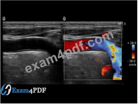
- A. Bidirectional
- B. Left to right
- C. Right to left
- D. Indeterminate
Answer: B
Explanation:
Comprehensive and Detailed Explanation From Exact Extract:
In the color Doppler image, the color bar indicates that red represents flow toward the transducer and blue represents flow away from the transducer. The vessel displays red flow on the left side and blue on the right side, indicating flow direction from left to right across the image.
According to sonography instrumentation reference:
"Color Doppler maps flow direction according to the color scale. Flow toward the transducer is coded as red, and away is coded as blue." Therefore, the correct answer is B: Left to right.
-
All answers are fully verified, 100% accurate, and aligned with Sonography Principles and Instrumentation documentation.
-
NEW QUESTION # 78
Which control should a sonographer use to change contrast resolution?
- A. Dynamic range
- B. Output power
- C. Gain
- D. Reject
Answer: A
Explanation:
* Reject: This control eliminates low-level noise and weak signals, affecting image quality but not primarily used for contrast resolution.
* Output Power: This adjusts the intensity of the transmitted ultrasound waves but does not directly change contrast resolution.
* Gain: This control amplifies all signals equally, affecting brightness but not specifically the contrast resolution.
* Dynamic Range: Adjusting the dynamic range changes the range of grayscale that the ultrasound system displays, which directly affects the contrast resolution by altering how many shades of gray are visible between the black and white extremes.
References:
"Understanding Ultrasound Physics" by Sidney K. Edelman
ARDMS Sonography Principles and Instrumentation study materials
NEW QUESTION # 79
Which setting can be increased to correct for clutter artifact when using pulsed-wave Doppler?
- A. Doppler gain
- B. Wall filter
- C. Sample volume
- D. Pulse repetition frequency (PRF)
Answer: B
Explanation:
The wall filter, also known as the high-pass filter, is used in Doppler ultrasound to remove low-frequency signals, which are typically associated with clutter artifacts. Clutter artifacts can be caused by tissue motion or vessel wall movement, and they appear as low-frequency signals that can obscure the desired blood flow signals. By increasing the wall filter setting, these low-frequency signals are filtered out, thus reducing the clutter artifact and providing a clearer depiction of the blood flow.
Reference:
American Registry for Diagnostic Medical Sonography (ARDMS). Sonography Principles and Instrumentation (SPI) Examination Review Guide.
NEW QUESTION # 80
What artifact is indicated by the arrows in the image below?
An ultrasound image of a fetus Description automatically generated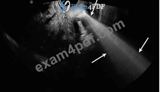
- A. Ring down
- B. Grating lobe
- C. Refraction
- D. Enhancement
Answer: A
Explanation:
Comprehensive and Detailed Explanation From Exact Extract:
The image shows a bright, continuous, vertical band extending from a gas-containing structure (seen at the top). This is characteristic of ring down artifact, which occurs when multiple small gas bubbles resonate and create continuous echoes below the structure.
According to sonography instrumentation reference:
"Ring down artifact results from resonance of gas bubbles, producing a continuous series of echoes distal to the source. It appears as a bright, vertical band that does not fade with depth." Therefore, the correct answer is A: Ring down.
-
NEW QUESTION # 81
What is an advantage of using pulsed wave Doppler as compared to using continuous wave Doppler?
- A. Decreased display of aliasing
- B. Higher echo sensitivity
- C. Ability to select sample depth
- D. Improved temporal resolution
Answer: C
Explanation:
Comprehensive and Detailed Explanation From Exact Extract:
The key advantage of pulsed wave Doppler is range resolution, meaning the operator can select a specific depth (sample volume) for measuring velocities. Continuous wave Doppler does not provide this capability, as it samples velocities along the entire beam path.
According to sonography instrumentation reference:
"Pulsed wave Doppler allows selection of sample volume depth, providing range resolution which continuous wave Doppler lacks." Therefore, the correct answer is B: Ability to select sample depth.
NEW QUESTION # 82
Which component of a contrast agent causes a marked mismatch in impedance between the agent and blood?
- A. Viscous liquid
- B. Solid
- C. Serous liquid
- D. Gas
Answer: D
Explanation:
Contrast agents used in ultrasound imaging typically consist of microbubbles filled with gas. The significant mismatch in acoustic impedance between the gas in the microbubbles and the surrounding blood creates strong reflectors of the ultrasound waves, enhancing the echogenicity of blood and improving the visibility of blood flow and tissue perfusion. The high contrast provided by the gas-filled microbubbles makes them particularly effective as ultrasound contrast agents.
Reference:
ARDMS Sonography Principles & Instrumentation Guidelines
Kremkau FW. Sonography Principles and Instruments. 9th ed. Philadelphia, PA: Elsevier; 2016.
NEW QUESTION # 83
Which factor does a string phantom evaluate?
- A. Flow velocity
- B. Slice thickness
- C. Two-dimensional resolution
- D. Intensity values
Answer: A
Explanation:
A string phantom is designed to evaluate the accuracy of Doppler ultrasound systems, specifically in measuring flow velocity. It consists of a moving string or filament that mimics blood flow within a vessel. By using this phantom, sonographers can assess how accurately the ultrasound system can detect and measure the speed of the moving target. This helps in calibrating and verifying the performance of Doppler systems, ensuring they provide accurate flow velocity readings in clinical practice.
American Registry for Diagnostic Medical Sonography (ARDMS) Sonography Principles and Instrumentation study materials.
Textbook of Diagnostic Sonography by Hagen-Ansert, S. L. (latest edition).
NEW QUESTION # 84
Which setting is the most likely cause of the artifact displayed in this image?
A close-up of a ultrasound Description automatically generated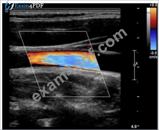
- A. Velocity scale set too low
- B. Color gain set too high
- C. Velocity scale set too high
- D. Color gain set too low
Answer: A
Explanation:
Comprehensive and Detailed Explanation From Exact Extract:
The image shows color aliasing, where the colors abruptly change within the vessel indicating wrap-around of Doppler shifts. This happens when the velocity scale (PRF) is set too low, causing velocities exceeding the Nyquist limit to alias.
According to sonography instrumentation reference:
"Aliasing occurs in color Doppler when the flow velocities exceed the Nyquist limit, commonly due to a low velocity scale (PRF)." Therefore, the correct answer is D: Velocity scale set too low.
-
NEW QUESTION # 85
Which spectral Doppler finding can occur when using a low pulse repetition frequency setting?
- A. Spectral broadening
- B. Dropout
- C. Range ambiguity
- D. Aliasing
Answer: D
Explanation:
Comprehensive and Detailed Explanation From Exact Extract:
Low PRF reduces the Nyquist limit, making the system more susceptible to aliasing when Doppler shift frequencies exceed this limit.
Principles and Instrumentation state:
"Aliasing occurs when Doppler shifts exceed half the PRF (Nyquist limit). Lowering PRF increases aliasing susceptibility."
* Spectral broadening (A) results from turbulence or poor angle.
* Range ambiguity (B) occurs at high PRF.
* Dropout (D) refers to signal loss, not aliasing.
Therefore, the correct answer is C: Aliasing.
-
NEW QUESTION # 86
Which can cause color aliasing?
- A. High wall filter
- B. High Doppler gain
- C. Low pulse repetition frequency
- D. Low frame rate
Answer: C
Explanation:
Comprehensive and Detailed Explanation From Exact Extract:
Color aliasing occurs when the Doppler shift exceeds the Nyquist limit, which is determined by the pulse repetition frequency (PRF). A low PRF reduces the Nyquist limit, making aliasing more likely.
According to sonography instrumentation reference:
"Aliasing occurs in color Doppler imaging when the Doppler shift frequency exceeds half of the PRF (Nyquist limit). A low PRF increases the likelihood of aliasing." Therefore, the correct answer is A: Low pulse repetition frequency.
-
NEW QUESTION # 87
Which resolution capability is most affected by spatial pulse length?
- A. Elevational
- B. Axial
- C. Temporal
- D. Lateral
Answer: B
Explanation:
* Axial resolution refers to the ability to distinguish two structures that are close to each other along the path of the ultrasound beam.
* Spatial pulse length (SPL) is the distance over which one pulse occurs, and it directly affects axial resolution.
* Shorter SPL improves axial resolution because it allows better differentiation of closely spaced structures.
* The axial resolution is improved by increasing the frequency of the transducer, which shortens the wavelength and hence the SPL.References:
* ARDMS Sonography Principles and Instrumentation guidelines on resolution parameters and their impact on image quality.
NEW QUESTION # 88
Which function is the purpose of the matching layer?
- A. Increase sound transmission
- B. Reduce ringing of the crystals
- C. Allow sequential firing of elements
- D. Convert acoustic to electrical energy
Answer: A
Explanation:
Comprehensive and Detailed Explanation From Exact Extract:
The matching layer is placed between the transducer's piezoelectric element and the patient's skin. Its primary function is to improve the transmission of sound energy into the body by reducing the acoustic impedance mismatch between the crystal and tissue, minimizing reflection at the transducer surface.
According to sonography instrumentation reference:
"The matching layer improves transmission of ultrasound energy by reducing impedance mismatch between the transducer and soft tissue, thus increasing the efficiency of sound energy transfer." Therefore, the correct answer is D: Increase sound transmission.
-
NEW QUESTION # 89
Which parameters determine the propagation speed of sound in a medium?
- A. Elasticity and density
- B. Frequency and impedance
- C. Intensity and density
- D. Amplitude and impedance
Answer: A
Explanation:
The propagation speed of sound in a medium is determined by the medium's elasticity and density. Elasticity refers to the ability of the medium to return to its original shape after deformation, while density is the mass per unit volume of the medium. The speed of sound increases with higher elasticity and decreases with higher density. This relationship is described by the equation#=##v=#E, where#vis the propagation speed,#Eis the elasticity (or modulus of elasticity), and##is the density.
References
* ARDMS Sonography Principles and Instrumentation (SPI) Exam Study Guide
* "Diagnostic Ultrasound: Principles and Instruments" by Frederick W. Kremkau
NEW QUESTION # 90
......
ARDMS SPI Exam Syllabus Topics:
| Topic | Details |
|---|---|
| Topic 1 |
|
| Topic 2 |
|
| Topic 3 |
|
| Topic 4 |
|
| Topic 5 |
|
Updated and Accurate SPI Questions for passing the exam Quickly: https://dumpstorrent.exam4pdf.com/SPI-dumps-torrent.html
